
Researchers have developed a new technique to map the complex microscopic interior of living cells in multiple colours simultaneously, overcoming a bottleneck in molecular biology. Researchers at the Jawaharlal Nehru Centre for Advanced Scientific Research (JNCASR) in Bengaluru designed a method called affinity encoding, which relies on synthetic, pumpkin-shaped molecular containers that hold onto cellular targets with varying degrees of grip. By selectively washing away and replacing fluorescent tags based on how tightly they cling to their targets, the team succeeded in painting internal cell structures in different fluorescent hues. The method even allowed them to create four-colour biological maps in live tissues.
To understand how biological systems work, scientists must observe multiple biomolecules working together inside a cell in real time. For years, they have relied on bioorthogonal chemistry. These are which are specialised chemical reactions, often described as molecular click chemistry, that snap fluorescent labels onto specific cellular parts without disturbing ordinary biological processes. However, researchers have hit a wall with this method. Setting up chemical reactions to colour cell structures inside living organisms without accidentally interfering with one another is extraordinarily difficult. As a result, scientists have largely been limited to observing only two cellular structures at a time, leaving most camera channels on modern fluorescence microscopes unused.
The team found an alternative solution by taking inspiration from nature. Instead of forcing cells to undergo a maze of distinct chemical reactions, the researchers decided to encode colour identity using differences in physical stickiness, or binding affinity. They turned to a technique called supramolecular host-guest chemistry, selecting a synthetic hollow molecule called cucurbit[7]uril, known simply as CB[7]. Shaped like a hollow barrel or miniature pumpkin, CB[7] acts as a host that can encapsulate smaller ‘guest’ molecules inside its cavity.
The breakthrough came from pairing CB[7] with two biocompatible guest molecules that bind to it with vastly different strengths. One guest, named 1-adamantylamine, binds to CB[7] with an exceptionally firm, ultra-high-affinity grip. The second guest, p-xylenediamine, binds securely enough for stable imaging, but roughly one million times more weakly than the first. This difference created a predictable hierarchy that the scientists could manipulate.
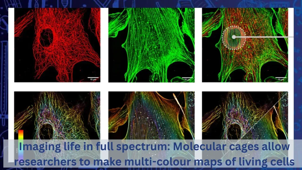
To demonstrate how the system works, the scientists attached the strong binder to cell scaffolds called microtubules and the weaker binder to fine structural strands called actin filaments. They first flooded the cells with CB[7] cages carrying a bright red fluorescent dye, causing both microtubules and actin to glow together in the same colour.
Next, they introduced a gentle wash containing free, untagged adamantylamine molecules. Because these free molecules were far hungrier for the CB[7] cages than the weak actin tags, they easily knocked the red cages off the actin, completely erasing its red glow in under three minutes. Meanwhile, the tightly bound cages on the microtubules refused to let go, remaining vibrantly red. Finally, the team introduced a fresh batch of CB[7] cages carrying a green dye. Because only the actin targets were vacant, they scooped up the green cages. In just a few sequential steps, a single type of molecular cage cleanly separated the two intertwined structures into distinct red and green channels without any spectral mixing.
The team confirmed this dynamic exchange worked across several increasingly complex test grounds. They validated the principle in test tubes, confirmed it inside cultured human cells, and successfully imaged thoracic muscle sections from adult fruit flies. Taking the experiment into dynamic living systems, they used specialised targeting drugs to label microtubules and actin inside the living gut tissues of fruit fly larvae, showing that displaced dyes were cleared harmlessly within thirty minutes without damaging the organism.
Going a step further, the researchers merged their two-tier host-guest system with two classic covalent click reactions. By combining affinity differences with chemical reactions, they captured stunning super-resolution images of human cells in four separate colours at once, clearly distinguishing actin, microtubules, energy-producing mitochondria, and sugary surface glycans.
This discovery could significantly improve our understanding of biological systems, medicine and modern healthcare. By breaking through the traditional two-colour barrier, this scalable imaging platform allows researchers to build far more comprehensive spatial maps of living tissues, dramatically improving our understanding of cellular machinery and accelerating the discovery of life-saving therapeutics.